Optomap Ultra-widefield Photography and Angiography
Regular detailed examination of the inside of the eye – the retina, is critical to eye health.
Doctors use a number of techniques to examine the retina including looking into the eye, usually after dilating and the use of special cameras for imaging inside the eye.
Until recently, most ophthalmic cameras could only photograph about 20% of the retina at a time. We now know that many eye diseases occur or begin at the outer edges of the retina, (“the periphery”), so examining this area is extremely important.
Because seeing the entire retina is so important at Shawnee Vision Source we have invested in the most advanced camera for ultra-widefield photography and angiography. In a single, quick shot, this camera produces “optomap” photos or angiograms of about 82% of the retina. These optomap images provide superior visibility of the retinal periphery allowing us to see, document, show you, and follow pathology that could not be seen with traditional eye cameras.
The optomap exam is quick and painless and combined with the thorough eye exams our doctors are trained to provide, this advanced technology offers a new level of diagnostic confidence.
We are proud and happy to offer this service to our patients. For more information please schedule an appointment with your eye care provider, and we will be in touch with you shortly.
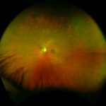
optomap Image showing healthy eye
optomap Image showing AMD
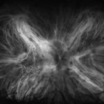
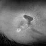
optomap Image showing Uveitis