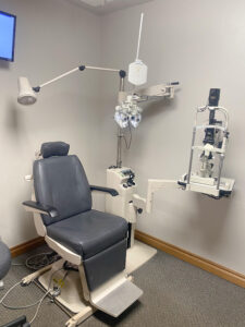
Comprehensive Eye Examination
The comprehensive eye exam at Shawnee Vision Source may require from half an hour to an hour. We will evaluate both your vision and the health of your eyes.
 Eye exams are vital to your overall health. Serious health issues like diabetes and high blood pressure are often first detected during an eye exam.
Don’t put it off — schedule your comprehensive eye exam at Shawnee Vision Source today.
Before you meet with the doctor, an ophthalmic technician will welcome you and perform an initial assessment of your vision with a series of preliminary tests. Upon reviewing your patient information, you may be asked several questions concerning your medical and vision history.
All or most of the following eye tests will be performed. Tests that are more specialized may be required depending on your condition.
Preliminary Testing Procedures
 Color Vision. The Ishihara Color Testing plates are used to determine the presence of red-green color deficiencies.
Depth Perception. A test to determine the presence of three-dimensional vision or stereopsis (binocular vision). Determines the presence of conditions such as amblyopia (lazy eye) or strabismus (cross-eyes).
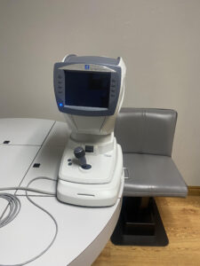
Auto Refraction. Computer controlled instrument used to provide an objective measurement of your refractive error or prescription for glasses or contact lenses. This quick, simple and painless test determines the depth and shape of the eyes by measuring how light is changed as it enters the eye.
Lensometry. This computerized measurement of your current eyeglass prescription allows the doctor to determine if there is a change in your prescription.
Soft Touch Tonometry (I care) Eye Pressure. This new devise tests the internal pressure of the eye for Glaucoma. The small instrument uses a light probe to briefly contact the cornea. This barely noticed measurement replaces the “Puff of Air” device.
Exam Procedures
 Visual acuity test. You will be asked to read the letters or numbers of an electronic eye chart projected on the wall. Progressively smaller letters are introduced as you read each line. Each eye is covered as the other eye is tested.
Eye Muscle Movement Test. To test muscle strength and control, the doctor will ask you to visually track a target in different directions and observe your eye movements.
Cover Test. This test will determine how well your eyes team together. As you focus at a small target some distance away, the doctor will cover and uncover each eye to observe how much your eyes move, watching for an eye that turns away from the target (strabismus). The test may be repeated with a target at close to you.
Confrontation Visual Field Exam. The examiner will ask the patient to cover one eye and stare at the examiner. The examiner will then move her hand out of the patient’s visual field and then bring it back in. The patient signals the examiner when her hand comes back into view. This is frequently done by an examiner as a simple and preliminary test.
External Exam and Pupillary Reactions. The doctor will observe the pupil reactions when a light and object is introduced at close distance. At the same time, the doctor will observe the exterior structures of your eye, examining for variations of the normal condition in the position of your eyelids and areas surrounding the eyes.
Retinoscopy. This test helps to establish your prescription. A streak of light will be directed into your eyes, as the examiner changes the lenses in an instrument (phoropter) in front of you. You will be asked to observe the letters at a distance through. Alternatively, an automated instrument (autorefractor) is used on most all patients for the same purpose.
Refraction Testing. The results of the computerized autorefractor are used as a starting point to refine your prescription. This is a series of questions, such as “Which is better, this or that?” while flipping back and forth between alternate lenses. Your prescription is better defined by selecting a preferred lens.
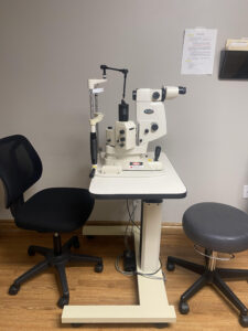
Slit-lamp (biomicroscope). This is a microscope, called a slit lamp, which magnifies and lights up the front of your eye. The doctor uses it to detect several eye diseases and disorders by examining each structure of your eye, including the cornea, iris, lens, and anterior chamber.
Retinal Examination (ophthalmoscopy). Using a head mounted light (binocular indirect) or an ophthalmoscope and pupil dilation, the doctor examines the inside structures of your eye, specifically the: retina, retinal blood vessels, vitreous, and optic nerve head.
Glaucoma Testing. This test may be performed as an alternate method to the Icare Tonometer test. It determines if the fluid pressure inside your eyes is within a normal range. Painless and taking just a few seconds, this test can be done several ways.
The Applanation Tonometer Test. This is another technique of accurately measuring the eye pressure. With drops numbing your eyes, a small devise is barely touched to the front surface of each eye with a glowing, bright-blue tool to measure the pressure.
Pupil Dilation (enlargement). With your pupils fully enlarged, the doctor will examine the inside of the eyes using various instruments and lights. The pupil enlarging drops for require approximately 20-30 minutes to take effect. The drops result in increased light sensitivity and blurred vision (especially at near) These effects may last for several hours or longer thus it is important to wear a sunglasses when leaving the office.
How often should I get a comprehensive eye exam?
Most eye care professionals recommend yearly eye exams. But it depends on your age, risk factors, and whether or not you wear corrective lenses.
The American Optometric Association (AOA) recommends children have their eyes examined at 6 months old, three years old, at the start of school, and every two years until age 18.
For adults, the AOA recommends a comprehensive eye exam every two years for ages 18 to 60 at minimum and annual exams for seniors age 61 and older.
What about contact lens exams?
A comprehensive eye exam typically does not include a contact lens fitting. So you may need to schedule a separate contact lens exam.
Just let us know when you schedule your appointment, and we’ll make sure you are taken care of.
Make healthy vision a priority in your life and in the lives of the ones you love. Schedule a comprehensive eye exam at Shawnee Vision Source now.